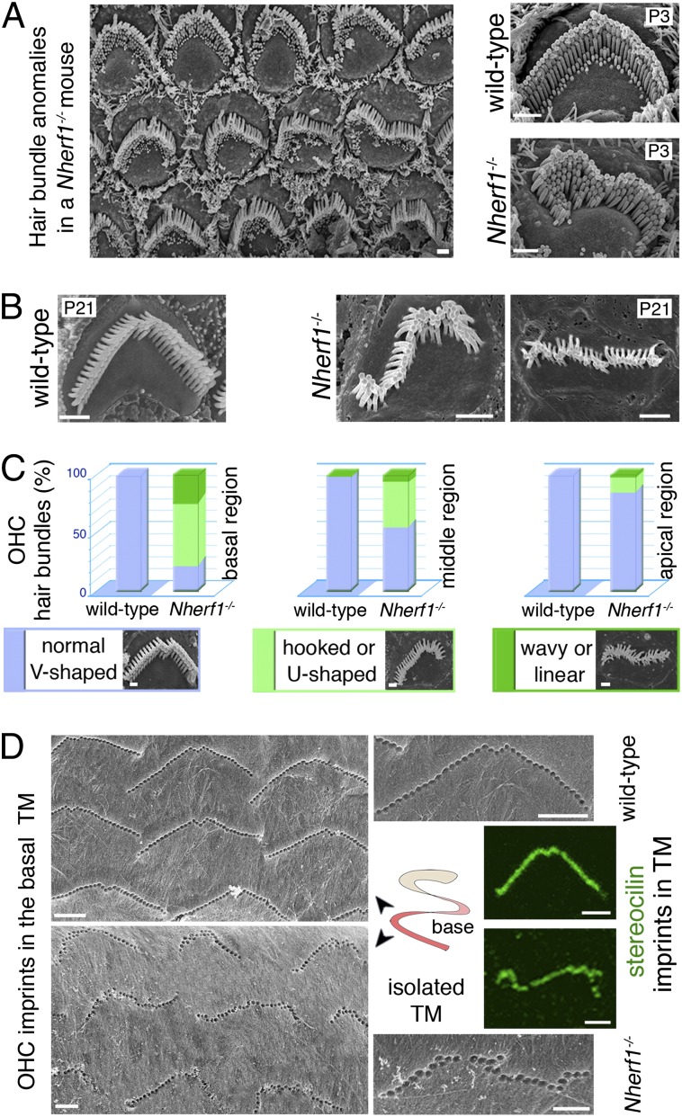
Fig. 2.
Abnormal OHC hair bundle shapes in Nherf1−/− mice. (A and B) In Nherf1−/− mice, the shapes of the OHC hair bundles are abnormal mainly in the basal cochlear region: wavy, linear, and hooked shapes are observed. (C) Abnormally shaped OHC hair bundles (light and dark greens, Lower) and normal, V-shaped hair bundles (blue) were counted in each cochlear region (basal, middle, apical) in Nherf1−/− and wild-type mice: in Nherf1−/− mice, 80 ± 5% of OHCs in the cochlear basal region displayed abnormal hair bundle shapes. (D) In Nherf1−/− mice (Lower), OHC imprints on the TM at the cochlear base (also labeled by anti-stereocilin antibodies, green) predominantly correspond to misshaped arrays of OHC stereocilia. (Scale bars, 1 µm.)

