Fig. 3.
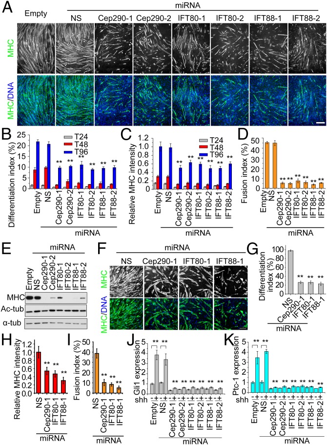
Inhibition of primary cilia assembly abolishes myoblast differentiation. (A) Inhibition of primary cilia in C2C12 cells suppresses differentiation. The differentiation efficiency of C2C12 cells expressing distinct miRNAs was examined at T24, T48, and T96. The empty and NS miRNAs were used as controls, and six miRNAs targeting Cep290, IFT80, and IFT88 were analyzed as indicated. Representative images of cells, visualized as indicated at T96, are shown. (B) Quantitative analysis of the differentiation index of the C2C12 cells expressing miRNAs at indicated time points. The differentiation index was calculated as the percentage of nuclei in MHC-positive cells. The differentiation indexes at T24, T48, and T96 are depicted by gray, red, and blue bars, respectively. Error bars, SD. (C) Quantification of the relative MHC intensity in each group as in A. Error bars, ±SD. (D) Quantification of fusion index at T96 as in A. Error bars, ±SD. (E) Western blots of extracts of C2C12 cells expressing individual miRNAs (Upper) and probed with indicated antibodies. (F) Inhibition of cilia assembly in primary mouse myoblasts suppresses differentiation. Differentiation efficiencies of primary mouse myoblasts expressing indicated miRNAs were examined by staining MHC (green) and DNA (blue). (G–I) Quantitative analysis of primary mouse myoblasts expressing different miRNAs shown in F. The differentiation index (G), relative MHC intensity (H), and fusion index (I) were measured. Error bars, ±SD. In each case, **P < 0.01. (J and K) Hh signaling is significantly diminished in C2C12 cells depleted of ciliary/centrosome components. The expression of Gli1 (J) and Patched-1 (K) was analyzed by qRT-PCR. Error bars, ±SEM. (Scale bars, 200 μm.)

