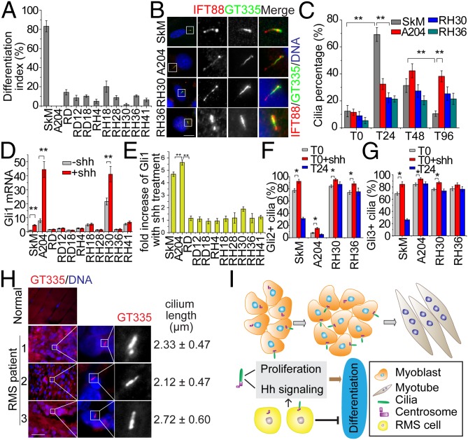
Fig. 4.
Ciliogenesis and Hh pathway are deregulated in RMS. (A) SkM and 10 RMS cell lines were induced to differentiate for 96 h, and the extent of differentiation indexes was assessed. Error bars, ±SD. (B) Representative images of primary cilia in SkM and three RMS cell lines. Magnified images of boxed Insets are shown on the right. (Scale bar, 10 μm.) (C) Quantification of cilia percentage at indicated stages of differentiation of SkM and RMS cells. Error bars, ±SD. (D and E) Analysis of Hh responsiveness in RMS cell lines. Gli1 expression was detected by qRT-PCR in SkM and RMS cells with or without Shh addition. The relative expression of Gli1 (D) and fold increase of Gli1 expression with Shh treatment (E) are shown. Error bars, ±SEM. (F and G) Quantitative analysis of the percentages of ciliated cells positive for Gli2 and Gli3 staining. (H) Staining of cilia and nuclei in normal muscle and in three RMS patients. Length of cilia is indicated for three patients. The lengths are indicated as mean ± SD. (Scale bar, 200 μm.) In each case, *P < 0.05; **P < 0.01. (I) The role of primary cilia in normal muscle differentiation and RMS. In myoblasts, primary cilia regulate cell proliferation and transduce Hh signaling. Dynamic assembly and loss of cilia occurs during myogenic differentiation, and this may limit the period of Hh signaling to a narrow window. See text for details.

