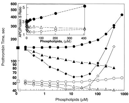
Figure 6.
Comparison of HDL anticoagulant potency with PL vesicles. The modified prothrombin-time assay was performed in the absence (open symbols) and presence (solid symbols) of added APC/protein S (see Methods) and varying concentrations of HDL (circles), PL vesicles containing 20% PS/80% PC (diamonds), or 1% PS/3% PE/96% PC (triangles). Squares indicate absence of added HDL or PL. Inset shows the ratio of the clotting time in the presence of added APC/protein S to that without added APC/protein S.

