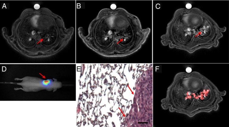
Fig. 1.
UTE MRI axial slices of the tumor-bearing mice (A) before and (B) after the i.v. administration of 200 μL of 50mM [Gd3+] USRP (day 35) or (C) the orotracheal administration of 50 μL of 50 mM [Gd3+] USRP (day 38). The presence and colocalization of the tumor was assessed with (D) BLI and (E) histology. In D the scale colors are proportional to the number of detected photons per second. The arrows indicate the tumor in the left lung. In E an HES stain (100× magnification) from an axial slice approximately corresponding to the MR image plane is shown. (Scale bar: 50 μm.) (F) Image showing the typical segmentation procedure used to contour the tumor. The red area delineates the tumor segmented from C. Vessels were recognized because of their hyperintense signal and typical shape, and were thus excluded from the segmentation procedure.

