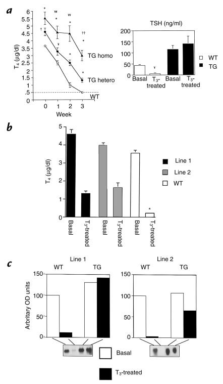
Figure 2.
Effect of T3 administration on serum T4, TSH concentrations, and pituitary TSH-β mRNA. (a) Total T4 concentrations (line 1) obtained at weekly intervals during the administration of pharmacological doses of T3 for 3 weeks. The hatched line represents the limit of detection of the T4 RIA. Mice with homozygous allelic expression of the transgene (TG homo; n = 5) were compared with TG mice harboring one transgenic allele (TG hetero; n = 13) and WT mice (n = 21). TSH concentrations were determined at baseline and at the end of 3 weeks in 11 heterozygous TG mice and 8 WT mice. Note that T4 and TSH are undetectable in WT mice after 3 weeks. In contrast, TG mice reveal only partial suppression of T4 and lack of suppression of TSH, indicating pituitary resistance. For the T4 data: ¥¥P < 0.01 and ††P< 0.0001, TG homo vs. TG hetero; ¥P < 0.01, †P< 0.001, *P < 0.0001, TG homo and hetero vs. WT. For TSH data: ¥P < 0.01, basal vs. treated WT. (b) Comparison of T4 concentrations before and after the administration of T3 in line 1 and line 2 TG mice. Note that despite basal thyroid hormone concentrations in line 2 that were similar to those of WT mice, T4 concentrations in line 2 are only partially suppressed to a level that is similar to that observed in line 1. TSH concentrations in line 2 mice were also partially suppressed to levels that were equivalent to untreated WT mice (44 ng/ml) after 3 weeks of T3 administration (data not shown). *P < 0.0001 compared with T3 treated values in lines 1 and 2. (c) TSH-β mRNA expression in pituitaries of T3 treated mice from both lines. Total RNA from pooled pituitaries (six mice per group) was analyzed by Northern blotting. The blots were initially hybridized with the mouse TSH-β cDNA and then with cyclophillin. Densitometry was used to determine mRNA abundance and cyclophyllin expression used to adjust the TSH-β message for differences in loading. TSH mRNA abundance is expressed in arbitrary OD units with WT basal expression normalized to 100. Note complete suppression of TSH-β message in WT mice, lack of suppression in line 1 mice, and partial suppression in line 2 mice. T3, triiodothyronine.

