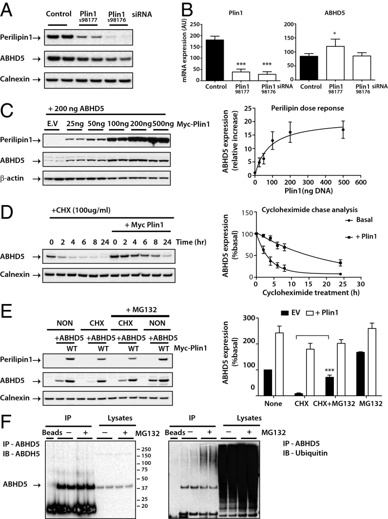
Fig. 2.
Perilipin 1 modulates endogenous ABHD5 protein expression through a ubiquitin-mediated proteosome degradation pathway. Differentiating 3T3-L1 adipocytes were transfected with control or two independent siRNA for perilipin 1 (Plin1). (A) Endogenous perilipin 1 and ABHD5 proteins were detected by immunoblotting and loading was assessed using an antibody to calnexin. Blots are representative of three separate experiments. (B) ABHD5 and Plin1 mRNA levels were determined by real-time PCR and expression normalized to mouse cyclophilin A. Values are mean ± SD of three independent experiments. *P < 0.05, ***P < 0.001. (C) Oleate-loaded COS-7 cells were cotransfected with Yn-ABHD5 and various amounts of Myc-perilipin 1. ABHD5 and perilipin protein expression was detected by immunoblotting and a representative blot is shown (Left) with the quantification of results from three independent experiments displayed (Right). (D) Oleate-loaded COS-7 cells cotransfected with Yn-ABHD5 and Myc-perilipin 1 (Plin1) were treated with 100 µg/mL cycloheximide, harvested at the times indicated and immunoblotted with antibodies to detect ABHD5 and perilipin 1 expression (Left). Quantification of ABHD5 expression from four separate experiments normalized to time 0 (basal) is displayed (Right). Note that in the presence of Plin1 the stability of ABHD5 expression is significant at all time points (P < 0.001). (E) COS-7 cells cotransfected with Yn-ABHD5 and Myc-perilipin were treated with cycloheximide (CHX) in the presence or absence of the protease inhibitor MG132 (10 µM) for 5 h. Cell extracts were immunoblotted with the antibodies indicated. Blots are representative of three separate experiments and quantification of these is shown on the right. Significance: ***P < 0.001 CHX vs. CHX+MG132. EV, empty vector. (F) Differentiated 3T3-L1 adipocytes were treated with MG132 (10 µM) for 5 h before cell lysis. ABHD5 was immunoprecipitated (IP) and the levels of ubiquitin detected by using a ubiquitin-specific antibody. Blots are a representative from two separate experiments.

