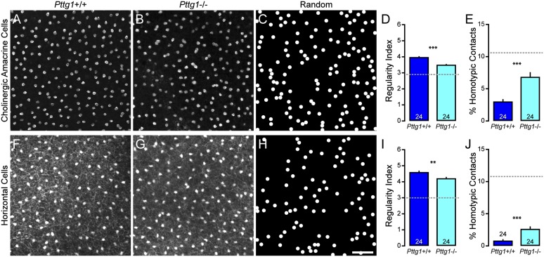
Fig. 4.
Cholinergic amacrine cell and horizontal cell mosaics are disrupted in Pttg1−/− mice. (A and B) Sample mosaics of cholinergic amacrine cells in the Pttg1+/+ retina (A) and Pttg1−/− retina (B). (C) Sample of a simulated random mosaic, matched in density and constrained by soma size. (D and E) Cholinergic amacrine cell mosaics in the KO retina had reduced regularity (D) and an increased frequency of homotypic pairs (E). (F–J) Similar results were found when analyzing the population of horizontal cells. n = number of fields analyzed, with the mean and SE indicated. The broken gray lines indicate the average value for random simulations matched in density and constrained by soma size. (Scale bar, 50 µm.) **P < 0.01 and ***P < 0.001.

