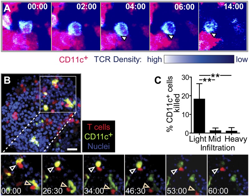
Fig. 4.
Stable T cell–APC interactions induce TCR signaling and APC killing in the islets. (A) TCR central supramolecular activating complex (cSMAC) formation and internalization during interactions with CD11c+ cells in the islets. OTI-TCR-GFP T cells were transferred into RIP-mOva.CD11c-mCherry recipients, and isolated islets were imaged by two-photon microscopy. TCR density is on a pseudocolor scale. Arrows indicate location of cSMAC, with internalized TCR visible in the last frame. Representative of two experiments. (B and C) APC killing by T cells in the islets. CD2-dsRed.OT-I T cells were transferred into RIP-mOva.CD11c-YFP recipients, and islets were imaged by two-photon microscopy. (B) Representative time-lapse of T cell-induced APC killing in a lightly infiltrated islet. Arrows indicate APC killing. Time-lapse represents 105 μm (x) × 105 μm (y) × 15 μm (z). Time stamp, min:sec. (C) Quantification of APC killing. Data combined from 10 experiments. **P < 0.01 by 1-way ANOVA Kruskal–Wallis test with Dunn’s Multiple Comparison Test.

