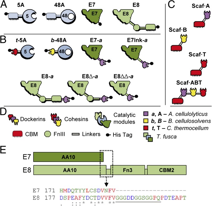
Fig. 1.
Recombinant proteins used in this study. Schematic diagram of the wild-type enzymes (A), chimeric enzymes (B), engineered scaffoldins (C), and key to the diagram (D). Each protein is color coded according to the source of the different modules, as follows: light green, dark green, and light blue, Thermobifida fusca; purple, Acetovibrio cellulolyticus; red, Clostridium thermocellum; yellow, Bacteroides cellulosovens. The numbers 5 and 48 refer to the corresponding CAZY family classification of the catalytic modules (GH5 and GH48). (E, Upper) Diagram of the modular architecture of E7 (Upper, dark green) and E8 (Lower, light green), to scale. (E, Lower) An excerpt of the amino acid sequence alignment (ClustalW) between E7 and E8 is provided, corresponding to the boxed section (dashed line) of the diagram. The linker segment is underlined. The color coding of the residues and the consensus symbols follow the standard ClustalW schemes.

