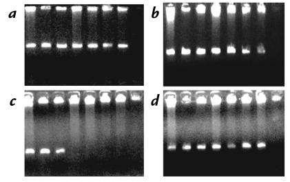
Figure 2.
Representative PCR analyses of the extent of the fibrinogen locus deletion. DNA samples in lanes 1–3 were from obligate carriers (individuals 3–5 in Fig. 1) and in lanes 4–7 were from patients (individuals 6–9). Lane 8 contains the negative control (no DNA). The amplified fragments were: (a) FGG exon 7; (b) FGA exon 1; (c) FGA exon 5; and (d) FGB exon 7. FGB, fibrinogen beta-chain gene; FGG, fibrinogen gamma-chain gene.

