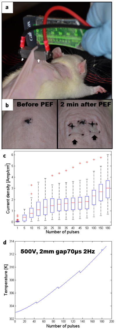
Figure 1.
Pulsed electric field ablation of rat skin. (a) Experimental setup for PEF procedure. Contact electrodes (arrows) are applied on the dorsal skin. (b) Morphological appearance of rat skin before the treatment (left panel) and 2 min after PEF treatment (right panel). Skin edema is marked with arrows. Ischemia at the electrodes points of contact is marked with stars. (c) Box plot of current densities (Amp cm−2) for each pulse (41 independent measurements per pulse are summarized). (d) Calculated skin temperature increase in Kelvin (K).

