Figure 3.
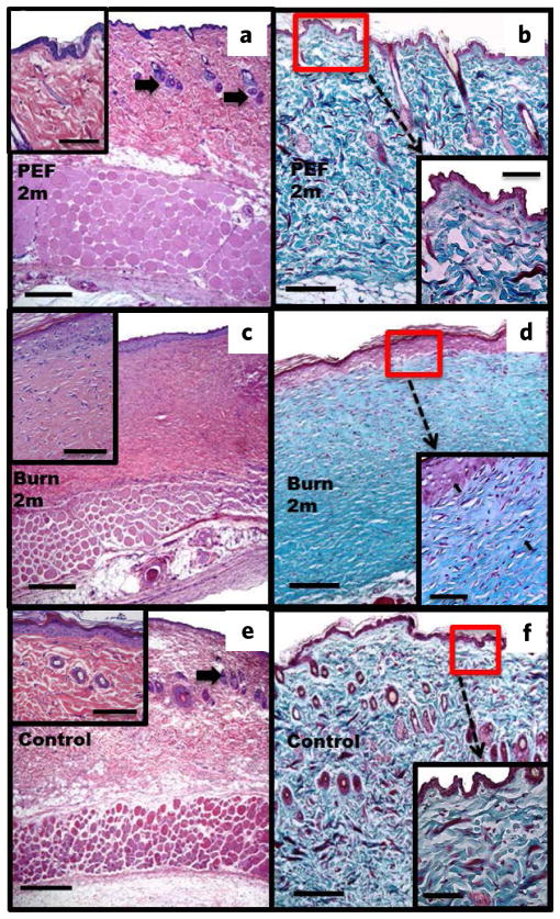
Histological analysis of rat skin after two months. (a,b) PEF injury. Shown is complete regeneration of structural and cellular skin components (a), Masson’s trichrome (b), complete restoration of epidermal spinous layer with 1–2 cell layers ((a), insert), hair follicles and sebaceous glands ((a) arrows). Collagen fiber structure is similar to the untreated skin ((b) vs (f) inserts). (c,d) Third-degree burn. Absence of skin structures such as hair follicles and sebaceous glands (c), Masson’s trichrome (d). Multicellular epidermis ((c), insert). The collagen fibers are parallel (arrows) and have a different structure compared to regular skin ((b) vs (f) inserts). (e,f) Untreated skin (e), Masson’s trichrome (f). Skin structures are shown with arrows (e). Normal-layered epidermis ((e), insert). Normal structure of collagen is shown in (f). Scale bars: (a,c,e) = 500 μm; (b,d,f) = 250 μm; inserts = 200 μm.

