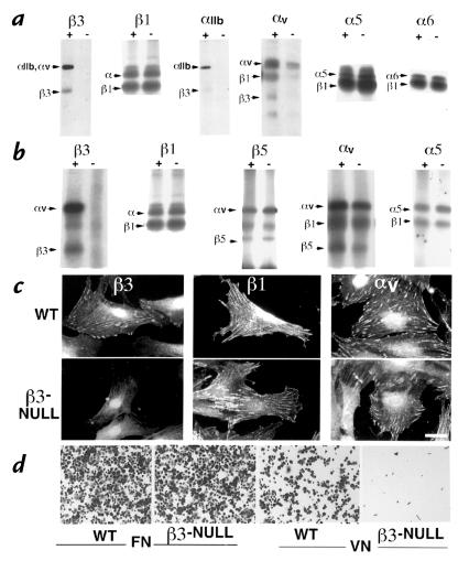
Figure 2.
Integrin profiles of cells isolated from the β3-null mice. (a) Surface iodination of platelets and immunoprecipitation of integrins β3, β1, αIIb, αv, α5 and α6 from wild-type (+) and β3-null (–) mice. (b) Surface iodination of mouse embryo fibroblasts (MEFs) and immunoprecipitation of β3, β1, β5, αv, and α5. (c) Immunofluorescence staining of β3, β1, and αv-integrins on wild-type and β3-null MEFs. (d) Adhesion assays with wild-type and β3-null MEFs on fibronectin and vitronectin. Bar (c), 15 μm.

