
Figure 7.
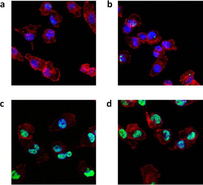
DNA damage study. Merge fluorescent images of Mia PaCa-2 cells 24 h after treatment with (a) 0.67 mg/mL Pd0-resins (negative control); (b) prodrug 5p (300 nM, negative control); (c) gemcitabine (300 nM, positive control); and 0.67 mg/mL of Pd0-resins + prodrug 5p (300 nM, BOOM activation assay). Fluorescent labels: Hoechst 33342 for cell nuclei (blue), Alexa Fluor 594 phalloidin for F-actin (red), and anti-phospho-histone γ-H2AX + Alexa Fluor 488 secondary antibody for phosphorylated γ-H2AX (green).