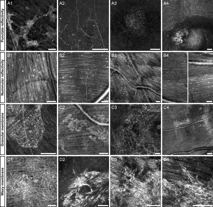
Figure 1.
Representative images of the first four features: punctate reflectivity (A1–4), nummular reflectivity (B1–4), granular membrane (C1–4), waxy reflectivity (D1–4). Diseases in each group include, rubella retinopathy (A1), achromatopsia (A2), optic disk pit (A3), normal (A4), normal (B1), glaucoma (B2), normal (B3), multiple sclerosis (B4), diabetic retinopathy (C1), Parkinson's (C2), branch retinal vein occlusion (C3), optic atrophy (C4), cone dystrophy (D1), central serous retinopathy (D2), birdshot choroidoretinopathy (D3), age-related macular degeneration (AMD, [D4]). (B4) is a contiguous vertical montage split into two halves, top is on the left and bottom is on the right. The first column of each row is highlighted below in Figures 3, 5, 7, 9. Scale bars: 100 μm.

