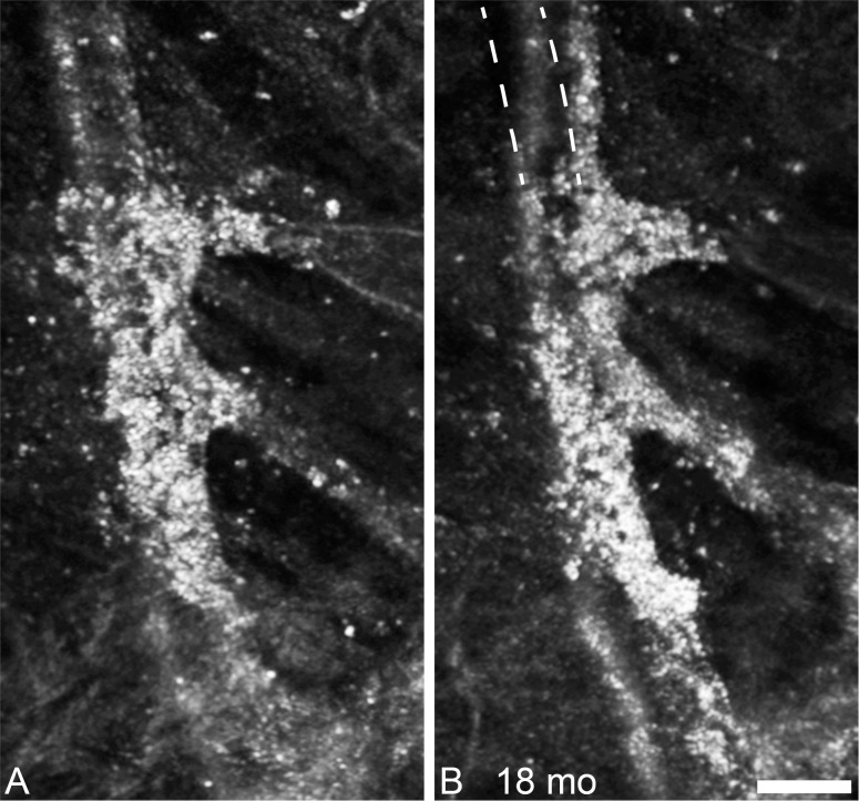
Figure 4.
Follow-up of punctate hyper-reflectivity, an enlarged portion of the lesion shown in Figure 3. In the 18 months between the first imaging session (A), and the second imaging session there are few, if any, punctate structures that have not changed position. The lesion appears to be enlarging along the vessels, and contracting, such that the vessel no longer follows its original course, as illustrated by the dashed lines in (B). Scale bar: 50 μm.

