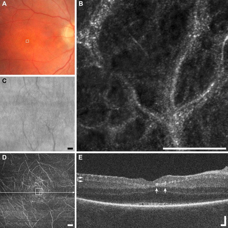
Figure 12.
Multimodal imaging of vessel-associated membrane example (E1), Leber's congenital amaurosis JC_0579. (A) Fundus photo appears normal. (B) An AOSLO image shows capillary loops entirely coated with a hyper-reflective membrane. Scale bar: 100 μm. (C) The SLO fundus image shows no obvious pathologic changes. Scale bar: 200 μm. (D) En face OCT segmented at the level of the GCL (horizontal arrows) shows hyper-reflective and disorganized vasculature. Scale bar: 200 μm. (E) The OCT B-scan shows many hyper-reflective spots (arrows), corresponding to hyper-reflective vessels. Scale bar: 100 μm.

