Figure 13.
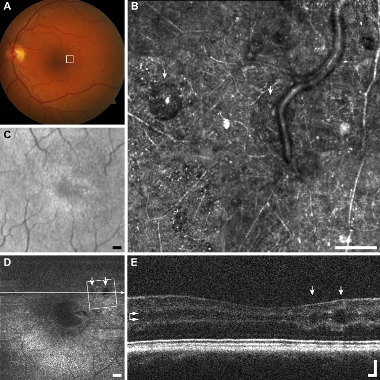
Multimodal imaging of microcysts example (F1), macular telangiectasia subject JC_10075. (A) Fundus photo does not resolve microcysts. (B) The AOSLO image shows a scattered distribution of very small (arrowheads) to very large microcysts (arrows). Scale bar: 100 μm. The borders of the cysts appear darker than the surrounding structure, and nearly all have a bright reflex on their apex. (C) The SLO fundus image shows disordered reflectivity, but no microcysts. Scale bar: 200 μm. (D) En face OCT segmented at the level of the INL (horizontal arrows) resolves only the largest microcysts (arrows). Scale bar: 200 μm. (E) The OCT B-scan shows the same two very large microcysts (arrows) seen in AOSLO imaging. Scale bar: 100 μm.

