
Figure 4.
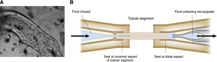
Study of isolated perfused tubular segments allowed study of each of the different nephron segments independently. (A) A photomicrograph of a portion of a rabbit proximal convoluted tubule during perfusion. (B) A schematic diagram of the technique. One end of the dissected tubule was connected to a micropipette, which was used to perfuse the lumen, and the other end was connected to a collection micropipette. Both the luminal fluid and the peritubular fluid could be controlled to assess tubular transport characteristics. A is reprinted with permission from Burg MB: Perfusion of isolated renal tubules. Yale J Biol Med 45: 321–326, 1972.