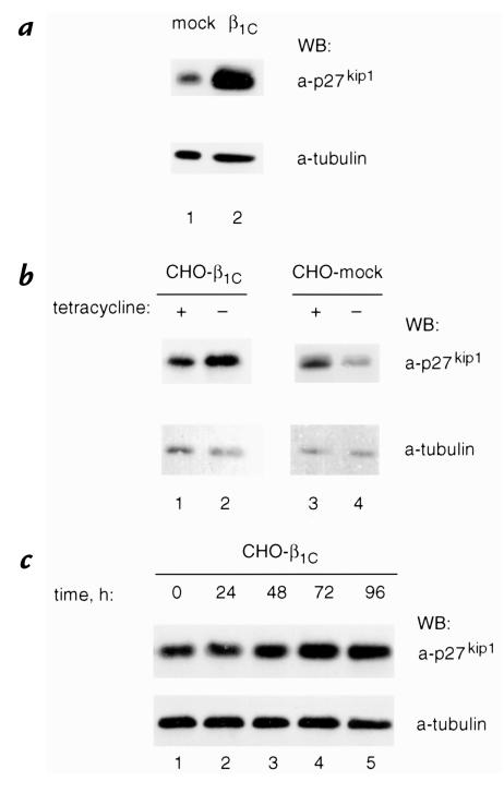
Figure 4.
β1C expression is accompanied by increased p27kip1 protein levels in NRP152 and CHO cells. (a) NRP152-β1C (lane 2) or NRP152-β1C-mock (lane 1) stable cell transfectants were cultured for 72 h in the absence of tetracycline, detached, and NRP152-β1C was seeded on tissue culture dishes coated with TS2/16, whereas NRP152-mock transfectants were seeded on tissue culture dishes coated with Ha2/5 for 1 h at 37°C, washed three times with serum-free medium, and cultured for 20 h in growth medium. Cells were then lysed and p27kip1 expression levels were evaluated by immunoblotting using 0.8 μg/ml MAB to p27kip1 (top). (b and c) CHO-β1C (b, lanes 1 and 2; c) or CHO-β1C-mock (b, lanes 3 and 4) stable cell transfectants were cultured for 72 h in b and for the indicated times in c, either in the absence (b, lanes 2 and 4; c) or in the presence (b, lanes 1 and 3) of 1 μg/ml tetracycline. In these experiments cells were not detached and allowed to reattach to MAB to β1 integrins, but were lysed in the tissue culture plate, and p27kip1 expression levels were evaluated as described in a. The experiments were repeated three times using two different β1C clones with similar results. Group differences were compared using Student's t test. The differences in p27kip1 expression levels in CHO-β1C, but not in CHO-mock transfectants in the presence or in the absence of tetracycline, are statistically significant (P = 0.03). Control for protein loading was provided by MAB to tubulin (a–c, bottom). Proteins were viewed by ECL. Time refers to the length of time in absence of tetracycline.

