
Figure 4. Increased apoptosis and reduced proliferation in SP8−/− facial mesenchyme.
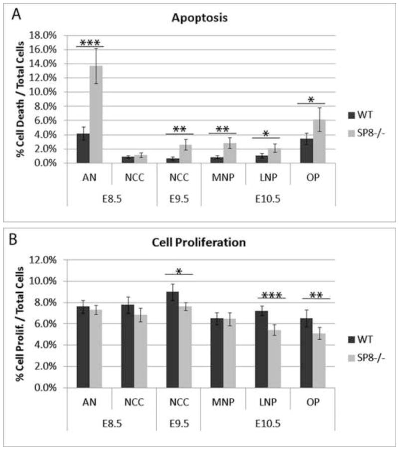
(A) Immunofluorescence for cleaved caspase 3 was used to label cells undergoing apoptosis. Wnt1-Cre activation of R26R-GFP labeled the neural crest. Apoptosis was quantified in various compartments of E8.5, E9.5, and E10.5 Sp8−/− and WT embryos. Results showed a significant increase in apoptosis in the anterior neuroepithelium at E8.5. The neural crest underwent elevated apoptosis at E9.5 as did the medial nasal prominence, lateral nasal prominence, and olfactory pit at E10.5.
(B) Immunofluorescence for phospho-histone H3 was used to label cells undergoing proliferation. The neural crest was labeled by Wnt1-Cre activation of R26R-GFP. Proliferation was quantified in various compartments of E8.5, E9.5, and E10.5 of Sp8−/− and WT embryos. Results showed a significant decrease in the neural crest at E9.5 as well as in the lateral nasal prominence and olfactory pit at E10.5.
Anterior Neuroepithelium, AN; Lateral Nasal Prominence, LNP; Medial Nasal Prominence, MNP; Neural Crest Cells, NCC; Olfactory Pit, OP
p<0.05 (*), p<0.01 (**), p<0.001 (***)