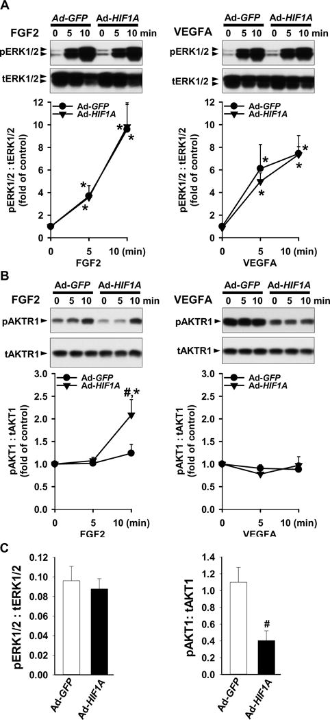
Fig. 3.

Effects of HIF1A overexpression on ERK1/2 and AKT1 phosphorylation in SCN-HUVECs. After infection with 100 MOI of Ad-GFP and Ad-HIF1A and 8 hr serum-starvation, cells were treated without or with 10 ng/ml of FGF2 or VEGFA under ~ 21% O2. Proteins were subjected to Western blot analysis and probed with specific antibodies against total (t) and phosphorylated (p) ERK1/2 (A) and AKT1 (B). Data normalized to tERK1/2 or tAKT1 are expressed as means ± SEM fold of the control. (C) After 2 days of transfection, basal levels of pERK1/2 and pAKT1 were analyzed by Western blot. Data are normalized to tERK1/2 or tAKT1 are expressed as means ± SEM fold of the control. *Differ from no growth factor control. #Differ from Ad-GFP at the same time point. p < 0.05, n = 4 cell preparations.
