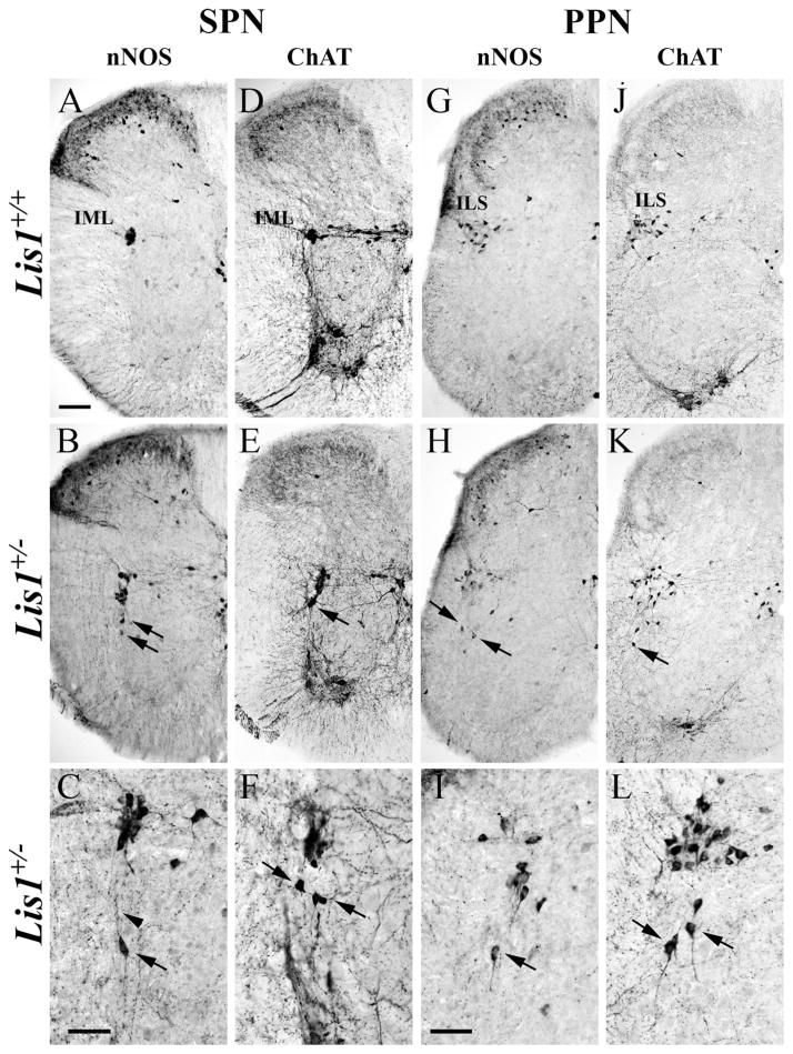
Figure 6.
Mispositioned sympathetic (SPNs) and parasympathetic (PPNs) preganglionic neurons persist in P30 Lis1+/− spinal cord sections and are identified with nNOS (A–C,G–I) and ChAT (D–F,J–L) antisera. A,D: In Lis1+/+ mice, nNOS- and ChAT-positive SPNs are found in the intermediolateral horn (IML). B–C,E–F: Lis1+/− SPNs in multiple sections are correctly positioned in the IML, but others are mispositioned ventrally (arrows) with dorsally oriented processes (C, arrowhead). G,J: In Lis1+/+ mice, nNOS- and ChAT-positive PPNs are concentrated in the intermediolateral sacral horn (ILS). H–I,K–L: Different sacral sections of Lis1+/− spinal cords illustrate incorrectly positioned PPNs (arrows) at lower (H,K) and higher (I,L) magnifications. Scale bar = 100 μm in A (applies to A,B,D,E,G,H,J,K); 50 μm in C (applies to C,F); 50 μm in I (applies to I,L).

