Figure 7.
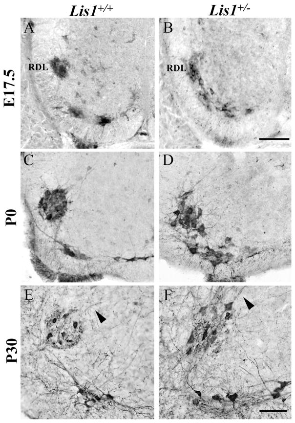
The retrodorsolateral group (RDL) of ChAT-positive somatic motor neurons (SMNs) is mispositioned in Lis1+/− lumbar spinal cord. A,B: On E17.5 the Lis1+/+ RDL nucleus (A) forms a tightly packed, circular group of neurons, but in Lis1+/− spinal cord (B) these cells are not yet separated from the ventral SMNs. C,D: At P0, the Lis1+/+ RDL nucleus is circular (C), whereas in the Lis1+/− RDL these cells remain loosely organized and close to ventral SMNs (D). E,F: At P30, the Lis1+/+ RDL nucleus is circular, with dorsomedially projecting dendrites (E, arrowhead). The Lis1+/− RDL nucleus is more oval-shaped, with dorsally directed dendrites (F, arrowhead). Scale bar = 100 μm in B (applies to A–D) and F (applies to E–F).

