
Figure 1.
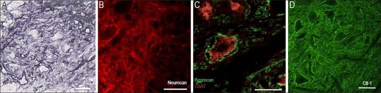
Examples of chondroitin-sulfated proteoglycan (CSPG) components in the ventral horn of the normal rodent spinal cord.
(A) Histochemical staining of transverse section from a mouse lumbar spinal cord with Wisteria Fluoribunda Lectin (WFA), which preferentially binds to carbohydrate structures terminating in N-acetylgalactosamine linked to galactose. The dark staining surrounding ventral neurons reveals strong CSPG-glycosaminoglycan content of the perineuronal nets as well as intercellular extracellular matrix (ECM) less tightly associated with the cell soma. (B) Immunostaining with monoclonal anti-neurocan antibodies show neurocan condensed around large neurons in the ventral horn of the intact rat spinal cord. (C) Aggrecan is identified using monoclonal antibody, Cat301. Dense aggrecan-rich perineuronal net structures surround a large motor neuron containing the cholinergic enzyme, choline acetyl transferase (ChAT; red). Note nearby motor neurons containing ChAT, but lacking aggrecan staining and small aggrecan-positive profiles which appear to be larger dendrites. (D) Immunostaining with antibody raised against the link protein Crtl-1 reveals proximity to most large neuronal cell bodies and loosely distributed throughout the gray matter in a rat spinal cord. Scales = 50 μm.