
Figure 1.
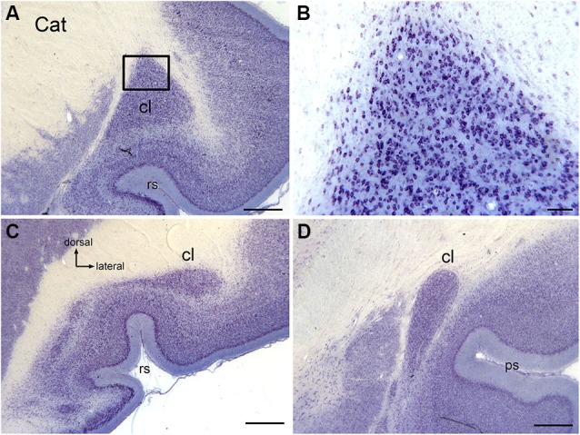
The claustrum in the cat shown on cresyl violet-stained celloidin-embedded sections. (A) The claustrum at about its rostro-caudal center. The rectangle shows the location of the image in (B). (B) Cells in the claustrum. (C) The claustrum at its rostral limit. (D) The claustrum at its caudal limit. Scale bars: A, C, D = 1 mm; B = 100 μm . Abbreviations: cl, claustrum; ps, pseudosylvian sulcus; rs, rhinal sulcus.