Figure 7.

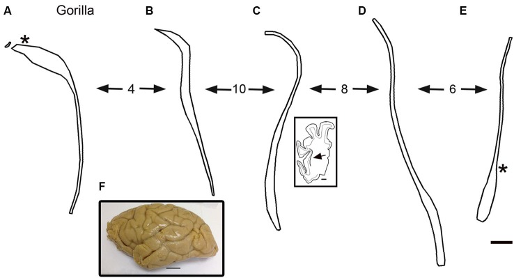
(A–E) Outline drawings of the claustrum of the gorilla from five coronal cresyl-violet stained sections. A is the most rostral. The spacing of the sections (mm) is indicated by the numbers between the arrows. (A) The asterisk shows the approximate location of the image in Figures 8A,C. The inset shows an outline drawing of the left half of the cerebral cortex on the section for the level of the claustrum shown; the arrow indicates the location of the claustrum on this section. (E) The asterisk shows the approximate location of the image in Figure 8C. (F) Lateral view of the left hemisphere of a gorilla showing the sulci and gyri. Caudal is to the right. Scale bars: E = 2 mm, same scale for A–D; C, inset = 5 mm; F = 2 cm.
