
Fig. 3. Activation of caspase-12 in the transgenic mouse.
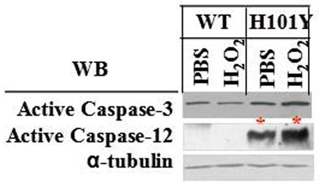
Four-week old mouse cerebellar slices in culture were treated with 250 μM H2O2 for 30 min. The samples were collected after treatments. The whole tissue extracts were used to determine active caspase-3 and caspase-12 by Western blotting. WT= control mouse cerebellar slice cultures; H101Y= PKCγ H101Y transgenic mouse slices in culture.