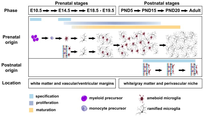
Figure 1.
Origin and development of microglia in the rodent CNS. The myeloid/mesenchymal-derived microglial progenitors start to colonize in neural tube around E10.5. Four days later, the second population of microglial progenitors originates from the circulating blood monocytes and/or fetal macrophages. The proliferating progenitors are differentiated and localized along vascular/ventricular margins and white matter during prenatal stages. Around PND5, these microglia are observed in both white matter and gray matter region, which are dramatically proliferate between PND5 and PND15. By PND20, the microglia are well matured with ramified morphology and stably distributed throughout the CNS. In the early postnatal and adult CNS, blood-borne precursors also generate a small number of perivascular ameboid-like macrophages/microglia.

