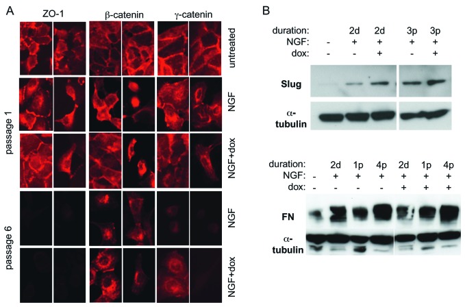
Figure 3.
Expression and localisation of EMT markers during the progression of c-erbB2-induced EMT. (A) Immunofluorescence micrographs of permeabilised TrE-ep5 cells stained with antibodies to ZO-1, β-catenin and γ-catenin. (B) Western blot analysis of the expression of EMT markers Slug and fibronectin (FN). Cells were treated with NGF or NGF + dox for the indicated durations (d, days; p, passages) or left untreated before lysis.

