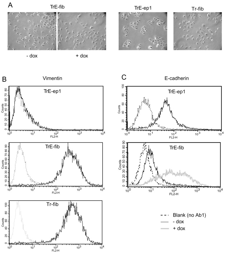
Figure 5.
Morphology and expression of vimentin and E-cadherin in the fibroblastic clone TrE-fib isolated after c-erbB2-induced EMT with concomitant induced expression of E-cadherin. (A) Micrographs showing morphology of TrE-fib cells with and without dox treatment for one week (prolonged treatment showed the same results). TrE-ep1 and Tr-fib cells (the latter generated by c-erbB2-induced EMT of Tr-ep cells, i. e. lacking the E-cadherin-IRES-GFP construct) (11,12), are shown for comparison. (B) Expression of the mesenchymal marker vimentin in TrE-ep1, TrE-fib and Tr-fib cells. (C) Expression of E-cadherin with and without dox treatment for two days in TrE-ep1 cells and TrE-fib cells. Expression levels in (B) and (C) were measured by flow cytometry (in B following permeabilisation by Triton X-100 treatment).

