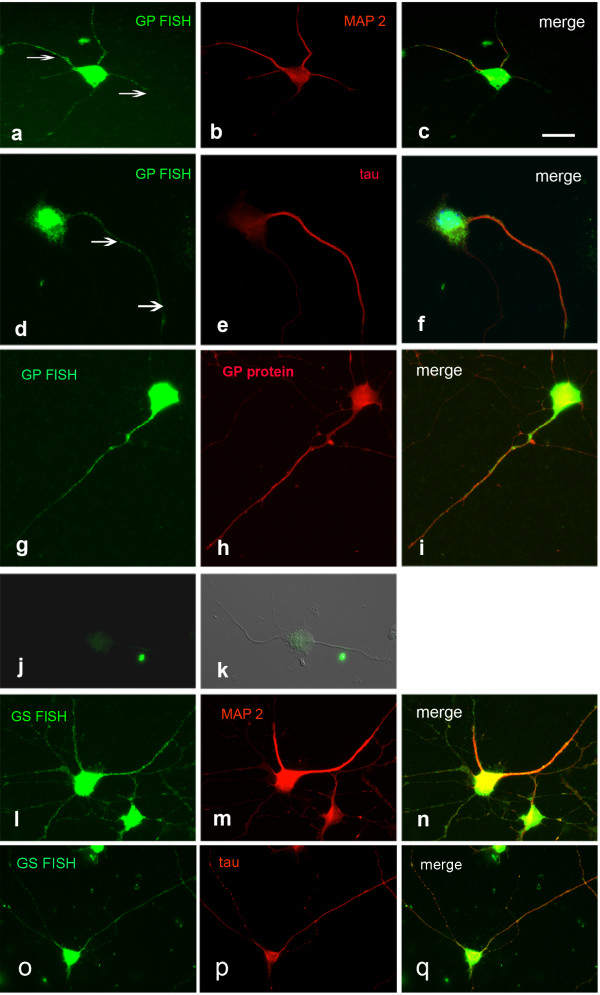
Figure 1.

FISH detection of GP and GS mRNA in spinal motoneurons, 5 DIV. GP mRNA colocalizes with MAP 2 (a-c) and tau (d-f). Arrows in a and d point to mRNA localized to neuronal processes. The signals for GP mRNA and protein are congruent (g-i). j, k Negative control applying GP sense probe instead of antisense. j FISH, k FISH combined with Nomarski optics. GS mRNA also colocalizes with MAP 2 (l-n) and tau (o-q). Bar in a = 20 μm and applies to a-q.
