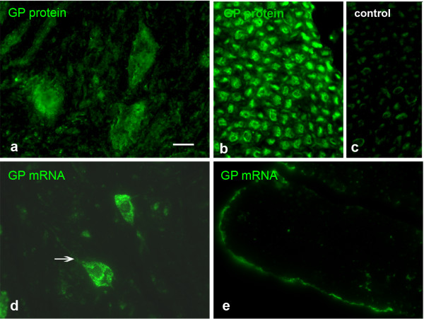
Figure 2.

GP IHC and FISH applied on sections of lumbar spinal cord and ventral spinal nerve. GP protein is prominent in large motoneurons (a) and the axons of the ventral root of the spinal nerve (b). Occasional signal can be detected in Schwann cells, but this is also the case in the negative control applying non-immune serum instead of immune serum (c). GP mRNA is mainly present in the neuronal cell bodies (d). No GP mRNA is present in the axons of the ventral spinal nerve (e). The arrow in d points to mRNA in the initial segment of a process. Bar in a = 20 μm and applies to a-e.
