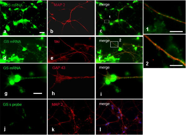
Figure 3.

FISH detection of GS mRNA in cortical neuronal cultures, 4 DIV. GS mRNA is prominent in neuronal cell bodies and colocalizes with MAP 2 in dendrites (a-c) and tau in axons. (d-f). Higher magnification (1 and 2) of the boxed areas in c and f reveals mRNA clusters in dendritic and axonal processes. FISH for GS mRNA combined with GAP 43 ICC reveals both signals in the axon, but colocalization in growth cones is not obvious (g-i). j-l Negative control applying sense probe in combination with MAP 2 ICC. Bar in a = 20 μm and applies to a-f and j-l. Bar in g = 10 μm and applies to g-i. Bar in 1 = 5 μm, bar in 2 = 10 μm.
