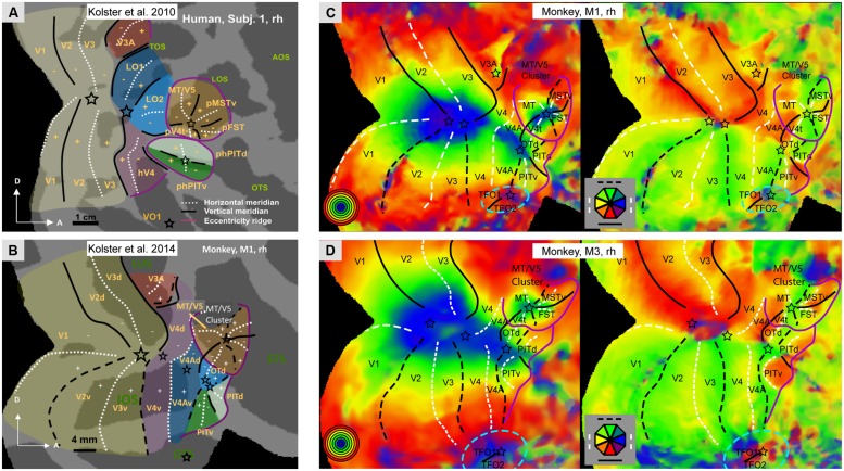
FIGURE 1.
(A,B) Schematic representation of the retinotopic organization of occipital cortex: in humans (A, subject 1, rh) and in monkeys (B, monkey M1, rh); Modified from Kolster et al. (2014).C,D: Polar angle and eccentricity maps for monkeys M1 (C) and M3 (D), same data as Janssens et al. (2014) but lower threshold. Black lines: vertical meridians (full: upper, dashed: lower), white dashed lines: horizontal meridians, stars: central visual field representation; purple lines: eccentricity ridges; In A,B: LuS: lunate sulcus, STS: superior temporal sulcus; OTS occipito-temporal sulcus; TOS: transverse occipital sulcus, LOS: lateral occipital sulcus, AOS: anterior occipital sulcus, OTS occipito-temporal sulcus; Other nomenclature: see Abbreviations. In C,D blue stippled elliptic outlines mark additional retinotopic regions (TFO1/2) ventral to V4A/PITv.

