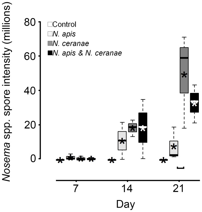
Figure 5. Nosema infection intensities in live-sampled adult worker western honey bees (Apis mellifera) at 7, 14, and 21 d post oral inoculation in control, Nosema apis, Nosema ceranae, and mixed N. apis/N. ceranae treatments.
Boxplots show interquartile range (box), median (black or white line within interquartile range), data range (dashed vertical lines), and outliers (open dots); asterisks (black or white) represent means. Horizontal square parenthesis under boxplots indicates a significant difference; controls were excluded from analyses because no infections were observed.

