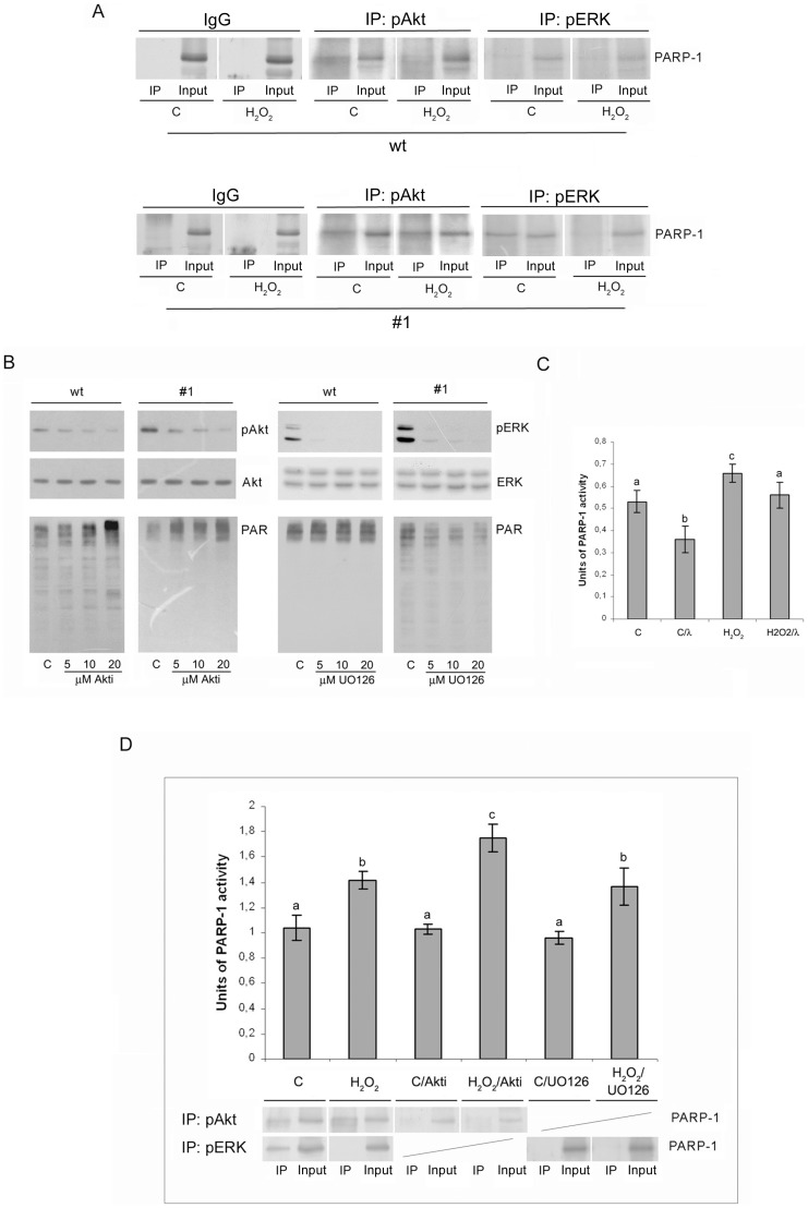
Figure 6. Modulation of PARP-1 activity mediated by Akt and ERK1/2 kinases.
(A) Immunoprecipitation of pAkt and pERK1/2 in lysates of control and hydrogen peroxide-treated wt and #1 cells. Immunoprecipitation was performed with anti-pAkt, anti-pERK1/2 and control IgG antibodies, and the obtained immunoprecipitates were probed with anti-PARP-1 antibody. (B) The effect of Akt inhibitor VIII (left panel) and MEK1/2 inhibitor (UO126) (right panel) on the level of poly(ADP-ribosyl)ation in control wt and #1 cells using immunoblot analysis with anti-PAR antibody (lower panel). The inhibition of kinases was estimated by immunoblot analysis with anti-pAkt, anti-pERK1/2, anti-Akt and anti-ERK1/2 antibodies (top panel). (C) PARP-1 activity assay performed with nuclear lysates from control and hydrogen peroxide-treated #1 cells and lysates that were dephosphorylated by lambda-phosphatase. (D) PARP-1 activity assay performed with nuclear lysates from control and hydrogen peroxide-treated #1 cells, without pretreatment or pretreated with Akt inhibitor VIII or MEK1/2 inhibitor (UO126). Every bar graph is accompanied by a corresponding immunoprecipitate; Immunoprecipitation was performed with anti-pAkt and/or anti-pERK1/2 antibodies, and the obtained immunoprecipitates were probed with anti-PARP-1 antibody. The results are expressed as means ± S.E.M. Means not sharing a common letter are significantly different between groups (p<0.05).

