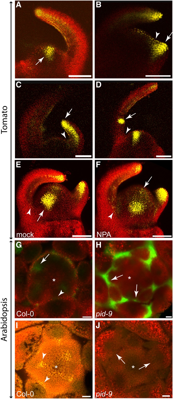
Figure 5.
Auxin Signaling in Tomato and Arabidopsis Shoot Apices Monitored by Confocal Microscopy.
(A) to (F) Shoot apices of 14-d-old transgenic tomato plants (cv M82) expressing DR5:VENUS. Auxin response is reflected by VENUS fluorescence (yellow).
(A) DR5:VENUS signals were detected at positions of the incipient leaf primordia (arrow).
(B) After bulging of a new leaf primordium, its adaxial side was labeled (arrow), whereas DR5:VENUS signals were reduced in the axillary region (arrowhead).
(C) During primordium outgrowth, DR5:VENUS signals were detected at the tip and in the provascular traces of the primordium (arrow). In the leaf axil, no DR5:VENUS signals could be detected (arrowhead).
(D) During leaf development, DR5:VENUS fluorescence was enhanced at positions where new leaflets were initiated (arrow). In the leaf axil, an auxin response was still not detectable at this stage (arrowhead).
(E) and (F) 3D reconstruction of mock- (E) and NPA-treated (F) DR5:VENUS tomato shoot apices. In mock-treated plants (E), DR5:VENUS signals were absent from leaf axils (arrowhead) but present in incipient primordia (arrow), as well as in tips and the provascular strands of developing leaf primordia. After NPA treatment (F), VENUS fluorescence could be detected in the axillary region (arrowhead) and was expanded in the SAM (arrow).
(G) and (H) Transverse sections through apices of transgenic Col-0 and pid-9 plants expressing DR5:GFP (28 d in SD).
(G) In Col-0, strong DR5:GFP fluorescence was monitored in the incipient leaf primordia (arrow). No GFP signal was detectable in the leaf axil (arrowhead).
(H) In pid-9, GFP fluorescence was detected in the axillary region of leaf primordia (arrows).
(I) and (J) Transverse sections through apices of transgenic Col-0 and pid-9 plants (28 d in SD) expressing DII-VENUS.
(I) In Col-0, DII-VENUS fluorescence signals were detected in axillary boundary zones (arrowheads), indicating low or no auxin levels in these regions.
(J) In pid-9, DII-VENUS fluorescence was not detectable in axillary regions (arrow), suggesting elevated auxin accumulation in these regions. Asterisks indicate the SAM.
Bars = 100 μm in (A) to (F) and 20 μm in (G) to (J).

