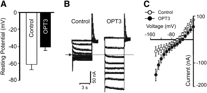
Figure 4.
OPT3 Is Functional in X. laevis Oocytes.
(A) Resting membrane potentials of OPT3-expressing (OPT3) and water-injected (Control) cells measured in standard ND96 recording solution.
(B) Example of OPT3-mediated currents (right panel) elicited in response to holding potentials ranging from 0 to −140 mV (shown only in 20-mV increments for clarity) in standard ND96 recording solution. Endogenous currents recorded in control cells are shown for reference on the left panel. The arrow and dotted line on the left margin indicates the zero current level.
(C) Mean current-voltage (I/V) curves constructed from the steady state currents recordings such as those shown in (B) for holding potentials ranging from 0 to −140 mV in 10-mV steps. Error bars represent se (n = 5).

