
Figure 2.
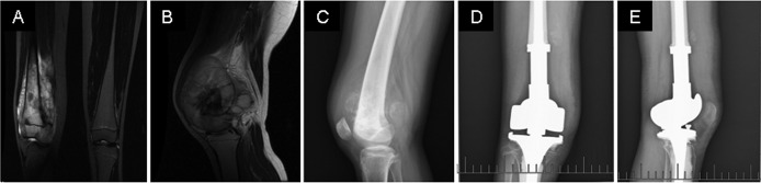
A patient who underwent knee arthroplasty (A and B) Preoperative MRI images show the tumor having crossed the physis. (C) A lateral X-ray image shows the tumor having crossed the physis. (D) An anteroposterior X-ray image shows the situation of the patient 6 months following knee joint and tumor resection, and reconstruction of the defect with metal prosthesis. (E) A lateral X-ray image shows the situation of the same patient 6 months following knee arthroplasty. MRI, magnetic resonance imaging.