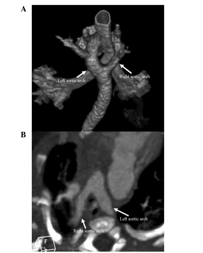
Figure 2.

A four-month-old male infant with DAA. (A) The CT volume-rendered image shows that the aortic arch split into two arches surrounding the trachea (thick left arch and small right arch). (B) An axial CT section image. The aortic arch split into two arches surrounding the trachea and esophagus. DAA, double aortic arch; CT, computed tomography.
