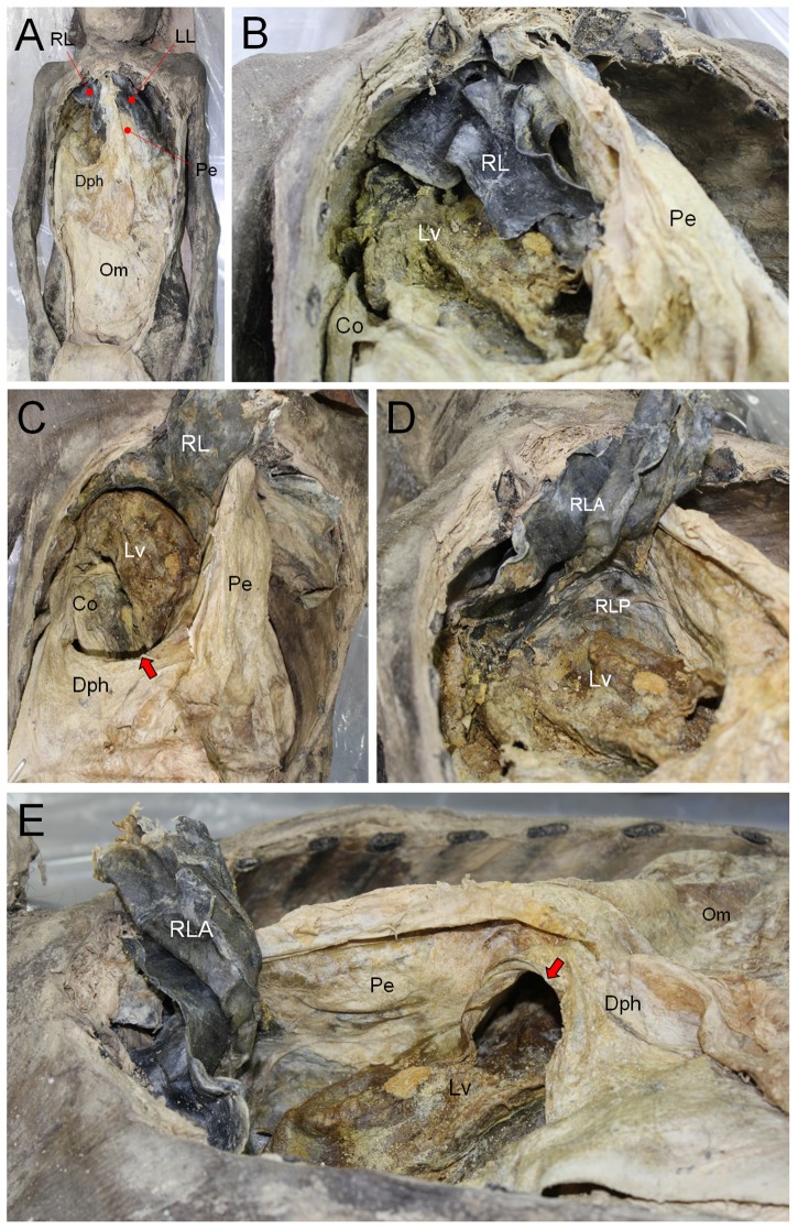
Figure 4. Dissection of the mummy.
(A) Thoracic and abdominal cavities exposed. Pe, pericardium; Dph, diaphragm; Om, omentum; Pe, pericardium; LL, Left lung; RL, right lung. (B) Magnified image of right thoracic cavity. Parts of liver (Lv) and colon (Co) could be found within the right thoracic cavity. (C) A part of RL is turned back. Lv and Co are much clearly identified. The defect in diaphragm is indicated by arrow. (D) Lv is protruding into the diaphragmatic surface of RL that is therefore folded into anterior (RLA) and posterior parts (RLP). (E) View from cephalic to rostral. Bochdalek hernia (indicated by arrow) in Dph is observed. Lv protrudes through the hernia defect.

