
FIG. 2.
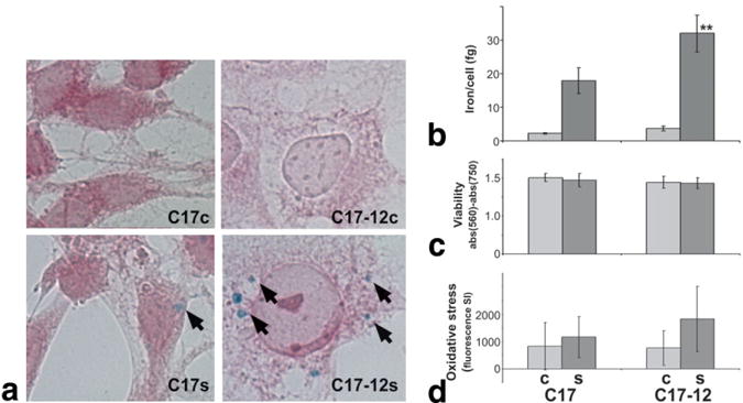
C17-12 cells accumulate significantly more iron than C17 control cells. a: Perls’ Prussian Blue stain for iron (arrows) with neutral red counterstain shows increased iron accumulation in supplemented C17-12 cells (C17-12s, lower right panel) compared to supplemented C17 cells (C17s, lower left panel). Cells grown in standard (control) medium (C17c and C17-12c) did not show iron accumulation by this method (top panels). b: AAS-quantified average iron content per cell in similar samples (**P < 0.05; N = 4). c: Viability of C17 and C17-12 cells under both control (light bars) and supplemented (dark bars) conditions was measured using the MTT assay (N = 8). d: CM-H2DCFDA reagent measurement of reactive oxygen species in all cell types is shown as total fluorescence minus autofluorescence (N = 8). Error bars represent the SD in each case.