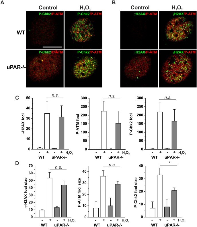
Figure 2. H2O2 induces DNA damage foci formation in WT and uPAR−/− mouse VSMC.
A. WT and uPAR−/− mouse VSMC were treated with H2O2 for 1 h, then fixed and stained for P-Chk-2 (Alexa 488) and P-ATM (Alexa 594). B. Cells treated as in C were stained for γH2AX (Alexa 488) and P-ATM (Alexa 594). Scale bar 10 µm. C. Quantification of H2O2-induced DNA damage foci number per cell nucleus was performed using Particle analysis tool of ImageJ. D. Average size of DNA damage foci was calculated using ImageJ.

