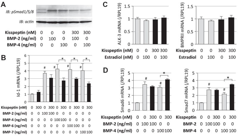
Fig. 3.

Effects of kisspeptin on BMP-Smad signaling in GT1-7 cells. (A) GT1-7 cells (1 × 10⁵ cells/ml) were precultured in serum-free conditions in the absence or presence of kisspeptin (300 nM) for 24 h, and then stimulated with BMP-2 and -4 (100 ng/ml). After 60-min culture with BMP treatment, cells were lysed and subjected to SDS–PAGE/immunoblotting (IB) analysis using anti-phospho-Smad1/5/8 (pSmad1/5/8) and anti-actin antibodies. The results shown are representative of those obtained from three independent experiments. (B) Cells (2 × 10⁵ cells/ml) were treated with BMP-2, -4, -6 and -7 (100 ng/ml) in combination with kisspeptin (300 nM) in serum-free conditions for 24 h. Total cellular RNA was extracted and Id-1 mRNA level was examined by real-time RT-PCR. The expression levels of target genes were standardized by RPL19 level in each sample. (C) Cells (2 × 10⁵ cells/ml) were treated with kisspepetin (300 nM) in combination with estradiol (100 nM) in serum-free conditions for 24 h. Total cellular RNA was extracted and ALK-3 and BMPRII mRNA levels were examined by real-time RT-PCR. D) Cells were treated with kisspeptin (300 nM) in the presence of BMP-2 and -4 (100 ng/ml) in serum-free conditions for 24 h. Total cellular RNA was extracted and Smad6 and Smad7 mRNA levels were examined by real-time RT-PCR. Results in all panels are shown as means ± SEM of data from at least three separate experiments, each performed with triplicate samples. The results were analyzed by ANOVA and, when a significant effect due to treatment was observed (P < 0.05), subsequent comparisons of group means were conducted. #P < 0.05 vs. control group; and * P < 0.05 between the indicated groups.
