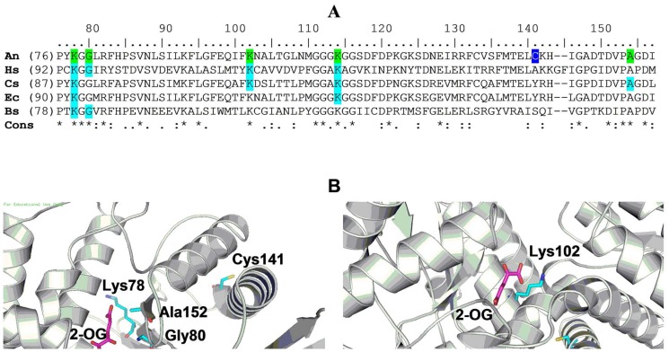
Figure 4. Location of Cys141 and Cys415 in the context of active site residues in AnGDH.
A) The GDH sequences (from UniProtKB database) from A. niger (An; B6V7E4), Homo sapiens (Hs; P00367), C. symbiosum (Cs; P24295), E. coli (Ec; P00370) and Bacillus subtilis (Bs; P39633) were compared (ClustalW). Residues implicated in glutamate binding (for GDHs where experimental evidence exists; shaded cyan) and the corresponding AnGDH residues (shaded green) are indicated. Cys141 is highlighted (in blue). B) AnGDH homology model showing Cys141 (left panel) and Cys415 (right panel) in the context of the enzyme active site. 2-OG: 2-Oxoglutarate.

