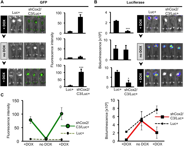
Figure 5. Reversible DOX-dependent suppression of Cox2-driven luciferase expression in skin of TPA-treated triple transgenic mice.
Luc+ mice and triple transgenic mice were subjected to the following diet, TPA skin application and imaging schedule: Mice were placed on a DOX-supplemented diet (+DOX) for 12 days followed by skin TPA application. Mice were imaged 24 hours later for GFP fluorescence and luciferase bioluminescence. The mice were then shifted to a DOX-free diet (no DOX) for 12 days and the skin TPA application, GFP fluorescence and luciferase bioluminescence analyses were repeated a second time. The mice were then shifted back to a DOX-supplemented diet (+DOX) for 12 days and the skin TPA application, GFP fluorescence and luciferase bioluminescence analyses were repeated a third time. (A) GFP fluorescence, indicating shCox2 expression. Data are means +/− S.D. (***p<0.001). After the DOX-diet was removed (middle panel, no DOX), GFP fluorescence returned to baseline (p>0.05, ns). (B) Luciferase bioluminescence, indicating Cox2 gene-driven luciferase expression. TPA-induced luciferase expression is reversibly reduced, in the presence of DOX, in triple transgenic mice. Data are means +/− S.D (*p<0.05, ***p<0.001). (C) Summary of GFP fluorescent intensity values (left panel) and bioluminescence (right panel) in Luc+ mice (dashed lines) and shCox2/C3/Luc+ mice (solid lines) for the three successive non-invasive imaging analyses. Error bars show S.D.

