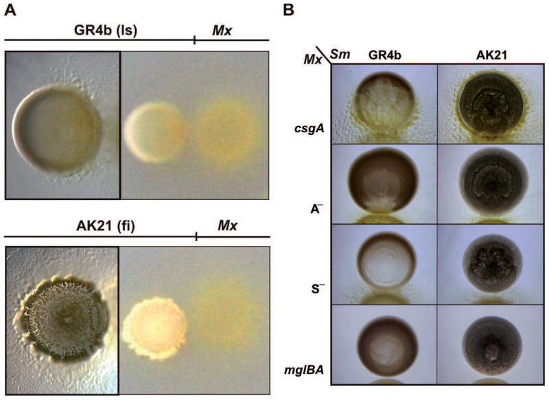
Fig. 1.

Predatory patterns of M. xanthus on S. meliloti. The non-mucoid reference laboratory strain (ls) GR4b and the mucoid S. meliloti field isolate (fi) AK21 were exposed to the M. xanthus (Mx) WT strain DK1622 (A) or mutants (B) impaired in rippling (csgA), A motility (A—), S motility (S—), and A— S— (mglBA) on CTT agar plates. Microscopic images of the S. meliloti-M. xanthus cocultures were taken 72 h after bacterial spotting. Images on the right of panel a were taken with a digital camera.
