
Figure 1. Mtbp is oncogenic.
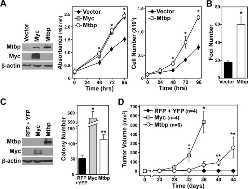
(A) Cell lysates of NIH3T3 cells expressing Flag-tagged Mtbp, Myc, or empty vector control were Western blotted. MTS assays (left) were performed, and viable cells were counted (right) at 24 hr intervals (*p<0.01 Myc or Mtbp vs. vector). (B) Cells described in A were placed in culture at low density and foci were quantified 7 days later (*p=0.0011). (C) Western blots and soft agar assays of NIH3T3 cells expressing RFP and YFP, Myc and RFP, or Mtbp and YFP; colonies quantified after 21 days (*p<0.0001, **p=0.0036). (D) Cells described in C were injected subcutaneously into nude mice at day 0, and tumor volume was measured at intervals (*<0.03 RFP+YFP vs. Myc, **p < 0.05 RFP+YFP vs. Mtbp). Number of mice indicated by n. Error bars are standard deviation (A–C)or standard error of the mean (D). P values determined by student’s t-tests.